
Microscope Accessories and Replacement Parts
General accessories and parts for use with microscopes. Products include lens oil, lens cleaner, slide dispensers, microscope covers, vessel holders, light cubes, and flow cells.
Many parts of your microscope can be updated or replaced including microscope lamps, lenses, and eyepieces. Replacement lamps, light sources, and power cords can extend the working lives of your microscopy equipment. New or replacement objectives and lenses can help increase the total magnification available with your microscope.
Add greater magnification or exchange your eyepiece for one with a reticule to more easily estimate the sizes of the items being viewed.
Replace or update your mechanical stage or clamps or add cell culture vessel adapters to facilitate specimen positioning.
Condensers can also be replaced, and conversion kits are available to add phase contrast, polarization, fluorescence, or other capabilities to your current system.
Some microscopes can be adapted for use with still or video cameras. Look for cameras and adapters designed for your specific microscope brand. Film and documentation-related products may also be available to support your record-keeping.
Temporary, semi-permanent, and more long-term slide storage options include:
- Slide racks that securely hold slides during staining procedures
- Folding fiberboard or plastic slide holders with horizontal indentations that keep microscope slides in place
- Slide storage boxes with vertical dividers; made from fiberboard, plastic, and wood and available in multiple sizes
- Slide holders and mailers for transporting slides safely
- Slide storage systems and cabinets for large quantities of slides or archival storage


Electron Microscopy Sciences Crossed Gauge Lines Microscope Graticule NE53, 19mm
Use as crossed lines, but for the measuring of distances between thelines. Greater accuracy can be obtained by locatng the specimen detail between the graticules gauge pair.
Size: 19mm
Divisions: Two vertical lines 0.1 mm apart


Electron Microscopy Sciences PremaDisk Diamond Grinding Disc SM Medium-Hard Bond, 15 µm Particles, 200 mm diameter
PremaDisk Flexible Diamond Grinding Discs offer superior surface finishes that are usually only seen in lapping processes.
The SM disc is a medium-hard resin bond base for fine grinding and polishing of metals, ceramics and glass.
The SH disc is a hard resin bond base for applications where a very sharp edge must be maintained on the workpiece.
Both discs increase productivity by combining optimum surface quality, removal rates, and tool life in the same process.
Particles: 15 µm
Diameter: 200 mm
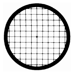
Electron Microscopy Sciences Veco Square Center Reference 150 mesh Copper Grid
Veco Grids offer superior handling characteristics. Plus, with a 0.8 mil thickness, Veco Grids are the most rigid grids available. 3.05 mm diameter.
Grid: Copper
Pitch: 167 µm
Hole: 117 µm
Bar: 50 µm
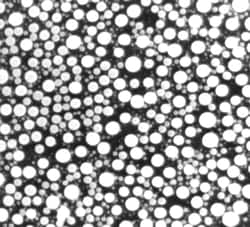
Electron Microscopy Sciences SEM Tin On Carbon Medium Resolution Test Specimen, JEOL Stub
Tin on Carbon is an alternate test specimen for medium resolution, and for the daily basic checking of your instrument performance. This specimen consists of a dispersion of tin spheres, within the size range 10 - 40 nm, on a carbon substrate. Ideal for astigmatism correction, it is also recommended for use in SEM's employed in the semi-conductor industry where the usual gold on carbon specimen cannot be used because of the risk of gold poisoning.
Type: JEOL Stub
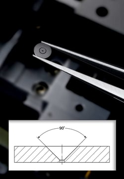
Electron Microscopy Sciences EMS Platinum Aperture with 300 µm hole, 0.12 mm thick
Platinum apertures are easily cleaned by either an aperture flamer or over a bunsen burner in the open air. We maintain a wide variety of Platinum Drilled Apertures in stock with different configurations suiting all of your needs. Nominal hole diameter: +/- 1 micron. Disc diameter: +/- 10 microns. Disc thickness: +/- 10 microns
Size: 300 µm
O.D.: 3 mm
Thickness: 0.12 mm

Electron Microscopy Sciences V-2 Quality, 25 x 25 mm, 0.15- 0.21 mm thick
EMS Mica Sheets offer a clean surface for EM app, carbon filming & particle spraying, as well as for AFM app. There are 2 types of mica: muscovite & phlogopite. Generally, one differs from the other by color (Muscovite is Ruby, Green or White; Phlogopite is Amber, Yellow, or Silver). The max operating temp for Muscovite is about 500-600 deg C & for Phlogopite is about 800-900 deg C. Our line consists of High Quality Muscovite Mica. This mica peels off very thin up to 0.0001" uniform layers, exposing "virgin" mica upon splitting.
Muscovite, potash type mica, sometimes known as granitic mica, is the best of all micas in dielectric strength, perfection of cleavage, & transparency. It has the chem formula: H2KAI3(Si04)3. Clear & Slightly Stained. Hard, uniform color, nearly flat & slight crystallographic discoloration. Very slight air inclusions & not more than 1/4 of the usable area.
Quality: V-2
Size: 25 x 25 mm
Thickness: 0.15- 0.21 mm

Electron Microscopy Sciences V-1 Quality, 25 x 75 mm, 0.26- 0.31 mm thick
EMS Mica Sheets offer a clean surface for EM applications, carbon filming and particle spraying, as well as for AFM applications. There are two types of mica: muscovite and phlogopite. Generally, one differs from the other by color (Muscovite is Ruby, Green or White; Phlogopite is Amber, Yellow, or Silver). The maximum operating temperature for Muscovite is about 500-600 degree C and for Phlogopite is about 800-900 degree C. Our line consists of High Quality Muscovite Mica. This mica peels off very thin up to 0.0001" uniform layers, exposing "virgin" mica upon splitting.
Muscovite, potash type mica, sometimes known as granitic mica, is the best of all micas in dielectric strength, perfection of cleavage, and transparency. It has the chemical formula: H2KAI3(Si04)3. Clear. Hard, of uniform color, nearly flat, free of all stains, foreign inclusion, cracks, and other similar defects.
Quality: V-1
Size: 25 x 75 mm
Thickness: 0.26- 0.31 mm

Electron Microscopy Sciences CoverWell™ Perfusion Chamber 8 well with 100 µl volume
CoverWell Perfusion Chambers allows up to 16 different specimens on one coverglass. "Press-to-Seal" covers form water-tight, multiwall cell incubation or cytochemistry chamber when pressed to coverslips or microscope slides. Reagents can be quickly added and removed through dual access ports (1.5 mm Dia) without disturbing or cross-contaminating specimens in adjacent wells. Chambers are removable for permanent mounting of specimens. CoverWell chambers are made from medical grade silicone bonded to clear, UV transparent plastic.
Volume: 100 µl

Electron Microscopy Sciences Veco Single Hole Grid A2000, Nickel with 2000 µm hole
Veco Grids offer superior handling characteristics. Plus, with a 0.8 mil thickness, Veco Grids are the most rigid grids available. 3.05 mm diameter .
Material: Nickel
Hole: 2000 µm

Electron Microscopy Sciences PremaDisk Diamond Grinding Disc SH Hard Bond, 15 µm Particles, 200 mm diameter
PremaDisk Flexible Diamond Grinding Discs offer superior surface finishes that are usually only seen in lapping processes.
The SM disc is a medium-hard resin bond base for fine grinding and polishing of metals, ceramics and glass.
The SH disc is a hard resin bond base for applications where a very sharp edge must be maintained on the workpiece.
Both discs increase productivity by combining optimum surface quality, removal rates, and tool life in the same process.
Particles: 15 µm
Diameter: 200 mm

Electron Microscopy Sciences CryoCapsule High Pressure Freezing
The CryoCapsule is used in High Pressure Freezing (HPF) and correlative light and electron microscopy (CLEM). Comparable to a small petri dish, it is composed of a landmarked sapphire disc and a gold spacer ring (50um thick) maintained together by a plastic ring. The specimens are encapsulated between the support sapphire disc (carbon landmarked) and a covering sapphire disc.
The CryoCapsule is loaded into a specific adaptor (HPM010, HPM100, HPF compact 02). Live cell imaging is done directly on the specimen in the CryoCapsule prior to HPF. Post-HPF, the specimen is processed for freeze substitution and room temperature sectioning.
CryoCapCell has developed a set of tools to manipulate the CryoCapsule in most scientific environments.
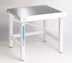
Electron Microscopy Sciences AMD-SM Microscope Workstation, 75 x 75 cm, Stainless Steel Surface
Microscope workstations featuring a unique Sorbothane isolator design removing up to 98 percent of unwanted vibrations.
Size: 75 x 75 cm
Surface: Stainless Steel
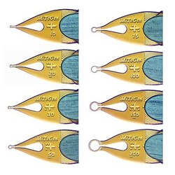
Electron Microscopy Sciences MicroMounts™ Crystal Tool with 150 µm Aperture and SPINE pin
MicroMounts are the high performance tools for manipulating and mounting protein crystals, virus crystals, and small molecule/inorganic crystals, and other small, fragile samples. They are ideal for both conventional and high-throughput X-ray crystallography applications.
Each consist of a thin (7.5um thick) micro-fabricated polyimide film, attached to a solid non-magnetic stainless steel pin. Film is wrapped around the steel pin and it provides excellent stiffness and convenient scoop-like action in retrieving and handling crystals.
Aperture: SPINE pin
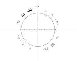
Electron Microscopy Sciences Walton & Beckett Graticule G24, 21 mm, 5:1 ratio
The Walton and Beckett graticule is for counting fibrous dust (asbestos or glass fibers) and particularly where the majority of fibers to be counted are shorter than 5 microns. The circle is divided into four by two diametrical lines scaled in units of 5 and 3 microns respectively. 3 and 5 microns are the critical measurements of fiber lengths and diameters used in fiber counting. Walton and Beckett include shapes with an aspect ratio of 3:1 or 5:1 for comparing to fibers.
IMPORTANT NOTE: The circle on these Walton & Beckett graticules must represent 100 microns at the stage and each one must be manufactured to the individual instrument. 5:1 ratio is useful where the majority of fibers to be counted are shorter than 5 um.
Size: 21 mm

Electron Microscopy Sciences TEM Screen Recoating RCA Viewing
For optimum viewing, fluorescent screens should be recoated regularly. Viewing screens are easily damaged by the electron beam, especially at high accelerating voltages where knock-on damage can be severe. A proprietary process where 1 to 2 micron particles of phosphor are deposited on the screen and produce the highest possible resolution.